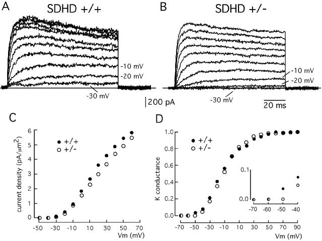
FIG. 5.
Macroscopic K+ currents in patch-clamped dispersed glomus cells of wild-type and partially SDHD-deficient mice. (A and B) Families of representative outward K+ currents in SDHD+/+ and SDHD+/− glomus cells recorded during 100-ms depolarizing pulses reaching membrane potentials between −30 and +60 mV in steps of 10 mV. (C) K+ current density (ordinate)-versus-voltage (abscissa) relationship in wild-type and SDHD+/− glomus cells. Each point is the average from at least eight different experiments. (D) Normalized K+ conductance (ordinate)-versus-voltage (abscissa) relationship in wild-type and SDHD+/− glomus cells. Each point is the average from at least three different experiments. Data points were fitted by an equation of the form G = 1/[1 + exp(V1/2 − Vm)/k]. The half activation (V1/2) (−11.7 and −10.6 mV for SDHD+/+ and SDHD+/−, respectively) and the slope factor (k) (14.6 and 12.6 mV for SDHD+/+ and SDHD+/−, respectively) were similar for the two curves. However, the activation threshold was clearly higher in SDHD+/− glomus cells (inset in panel D). In panels C and D, error bars are omitted.

