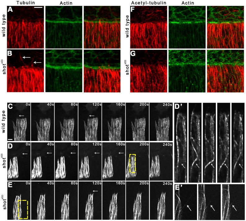
Fig. 3.
Abnormal MTs in the shotsf20mutant DME cells. (A,B) Immunofluorescence staining of wild-type (A) and shotsf20 mutant (B) embryos at the dorsal closure stage. Tubulin staining labels the MT network (red); actin is labeled with phalloidin (green). White arrows indicate abnormally long and curled MTs at the leading edge of shotsf20mutant DME cells. (C,D) Frames taken from movies showing Tubulin–EGFP-expressing DME cells in wild-type (C) and in shotsf20mutant (D,E) embryos. White arrows indicate MTs growing into protrusions. MTs are abnormally long and curled at the leading edge of shotsf20mutant DME cells. (D′,E′) Enlargement of the boxed regions in D and E showing bending of a MT in D′ and protrusion of a MT at the lateral surface in E′. (F,G) Staining for acetylated tubulin labels stabilized MTs (red); actin is labeled with phalloidin (green). In shotsf20 mutants, no abnormal distribution of stabilized MTs is detectable. Scale bars: 5 µm (C,D,E); 10 µm (A,B,F,G).

