Fig. 1.
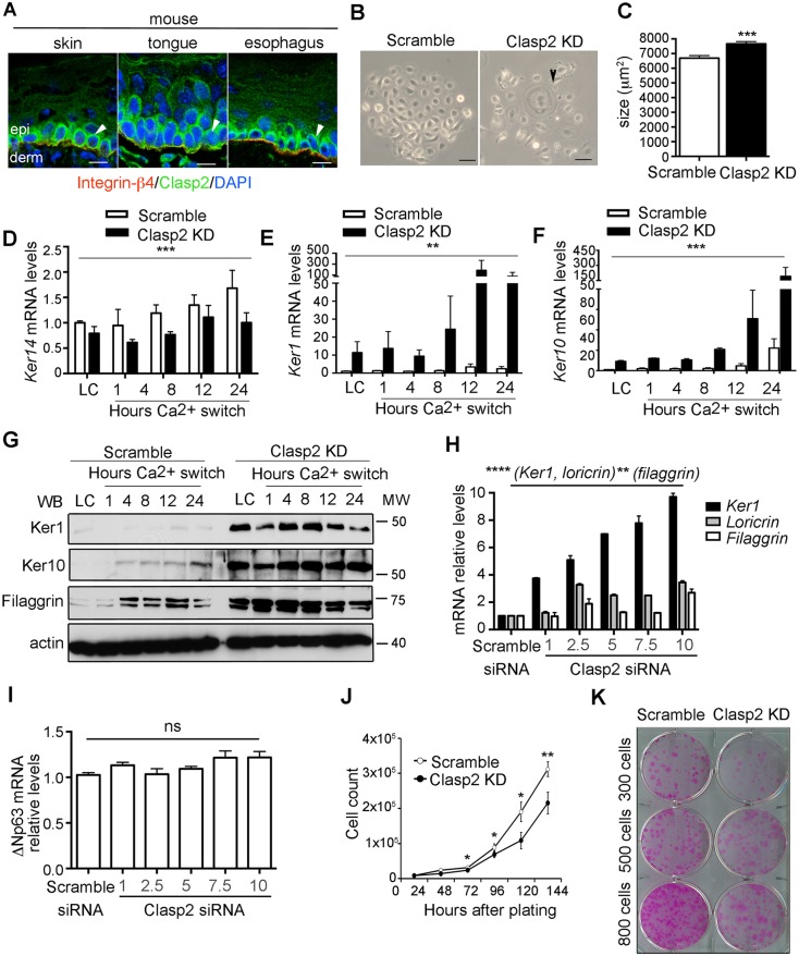
Clasp2 expression in mouse keratinocytes prevents premature differentiation. (A) Clasp2 localization in stratified epithelia (arrowhead). Epi, epidermis; derm, dermis. Scale bars: 10 μm. (B) Scramble and Clasp2KD mouse keratinocytes brightfield images. Arrowhead indicates a differentiated cell. Scale bars: 100 μm. (C) Quantification of cell size (n=562 scramble and 645 Clasp2KD cells). (D) Ker14 mRNA levels in scramble and Clasp2KD mouse keratinocytes relative to levels of Gapdh. Hours Ca switch, time after Ca2+ switch. (E,F) Ker1 and Ker10 mRNA levels relative to that of Gapdh at different time points after Ca2+ addition. LC, low Ca2+. (G) Ker1, Ker10 and filaggrin immunoblots. (H) mRNA levels of differentiation genes relative to that of actin and (I) mRNA levels of ΔNp63 in scramble and mouse keratinocytes that had been treated with different concentrations (μM) of siRNAs against Clasp2 (Clasp2 siRNA). (J) Proliferation curves of scramble and Clasp2KD mouse keratinocytes. (K) Colony formation assay. Data are presented as mean±s.e.m. *P<0.02, **P<0.01, ***P<0.002 (C) Mann–Whitney U test, (D) two-way ANOVA test, (E,F) Kruskal–Wallis test, (H,I) one-way ANOVA test, (J) two-tailed Student's t-test; ns, non-significant. n=2–3 independent experiments per panel.

