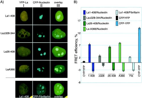
FIG. 5.
FRET analysis reveals SBM-dependent interaction of La with nucleolin in the nucleolus. (A) Constructs used for the interaction analysis below were first examined for expression in HeLa cells prior to FRET analysis. Column I shows the yellow channel fluorescence of the YFP-hLa constructs, column II shows the cyan channel fluorescence of the CFP-nucleolin constructs, and column III shows the overlaid views for the indicated constructs. Note that the last row shows CFP-fibrillarin. (B) Acceptor photobleaching was used to monitor FRET between the YFP-hLa (acceptor) constructs and the CFP-nucleolin or CFP-fibrillarin (donor) after specific bleaching of the acceptor with YFP-wavelength light. FRET efficiencies reflective of positive interactions were plotted as bars above the horizontal line (see Materials and Methods). Bars below the horizontal line reflect control measurements in the same cells in nucleolar areas in which there was no prebleaching of the acceptor. At least 30 measurements were collected and quantitated for each bar in the graph. CFP/YFP, CFP alone plus YFP alone; CFP-YFP, CFP-YFP fusion protein.

