Figure 1.

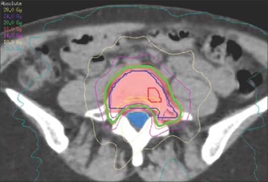
Axial CT scan demonstrating dosimetry for a lumbar spine lesion treated with VMAT, highlighting the steep dose gradient generated between the involved vertebral body and the thecal sac (blue). Adapted with permission[19]

Axial CT scan demonstrating dosimetry for a lumbar spine lesion treated with VMAT, highlighting the steep dose gradient generated between the involved vertebral body and the thecal sac (blue). Adapted with permission[19]