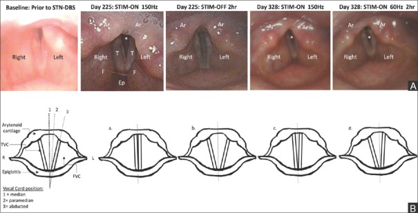
Figure 2.
(A) Laryngoscopic images of laryngeal airway opening. T = True vocal folds; F = False vocal folds; Ep = Epiglottis; Ar = Arytenoids; * = Airway (space between vocal folds). (B) Schematic representation of vocal fold mobility. (a) True vocal fold adduction during phonation. (b) Vocal fold abduction during normal inspiration. (c) Vocal folds immobile at the paramedian position. (d) The left vocal fold appears immobile at the paramedian position while the right vocal fold is abducted during inspiration. TVC = True vocal cords/folds; FVC = False vocal cords/folds

