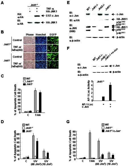
FIG. 6.
Distinct roles of JNK1 and c-Jun in apoptosis. (A) Jnk1−/− cells were transfected with an expression vector encoding HA-JNK1 or empty vector (3 μg each). The activity and expression of HA-JNK1 were measured as previously described (18). An in vitro kinase assay (KA) and immunoblotting (IB) were performed with anti-HA antibody (α-HA). (B) Jnk1 null cells were transfected with an expression vector encoding EGFP (0.8 μg) and HA-JNK1 or empty vector (3.2 μg each) for 12 h, and then the cells were infected with Ad/IκBα(AA) for 16 h. Cells were treated with TNF-α (5 ng/ml) for 6 h or left alone (control). Apoptosis of EGFP-positive cells was measured as described previously (26). The cells were examined by phase-contrast microscopy or by microscopy with Hoechst 33258 staining. (C) Schematic presentation of the results in panel B. C, control; T/AA, TNF-α (5 ng/ml) plus Ad/IκBα(AA). (D) As in panel B, except the cells were irradiated with UV (60 or 10 J/m2) for 24 h. (E) Jnk1 null cells were transfected with expression vectors encoding HA-JNK1 or empty vector (3 μg each), and c-Jun expression was measuredby immunoblotting analysis using anti-c-Jun antibody. Expression of HA-JNK1 and different isoforms of JNK was measured by immunoblotting analysis using anti-HA antibody and anti-JNK antibody (antibody 666; PharMingen), respectively. (F) Jnk1 null cells were transfected with expression vectors encoding the AP-1 Luc reporter gene (0.5 μg), along with c-Jun or empty vector (3 μg each). The levels of transfected c-Jun protein expressed in Jnk1−/− cells were analyzed by immunoblotting using anti-c-Jun antibody. WT, Jnk1−/−, and Jnk2−/− cells were used as controls. AP-1 Luc activity was measured as described previously (17). (G) Jnk1 null cells were transfected with expression vectors encoding EGFP (0.8 μg) and c-Jun or empty vector (3.2 μg each). The cells were then treated with TNF-α (5 ng/ml) for 6 h plus Ad/IκBα(AA) (T/AA) or with UV (60 or 10 J/m2) for 24 h or left alone (control [C]). Apoptosis was analyzed as described in the legend to Fig. 5A.

