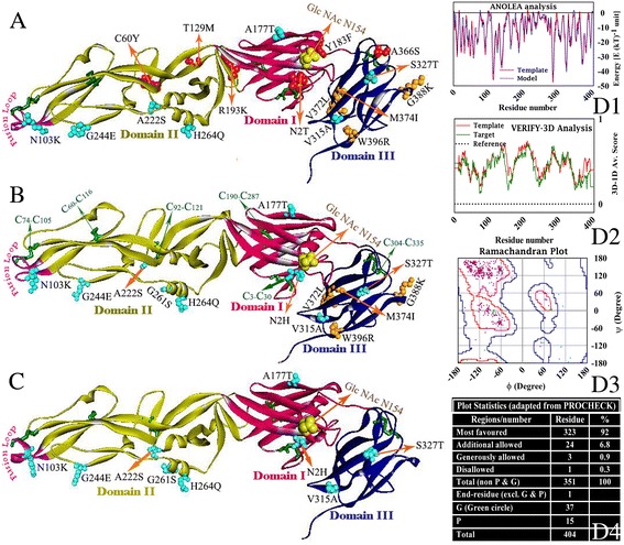
Fig. 3.

Backbone Cα-traces of homology model (a for Ishikawa, b for JEV45 and c for JEV28) of the ecto domain of PM0080325, PM0080324 and PM0080323 respectively. Domain I (red), domain II (yellow) and domain III (blue) are shown in different colors. Fusion loop (purple) and substitutions with respect to the template (SA14-14-2) are highlighted in each of the model based on their occurrence (see Table 1). Disulfide bonds (green) are also highlighted in each of the model based on their occurrence and only labeled in case of JEV45. Comparison of ANOLEA-profiles [61] of PM0080324 (green trace) with template (red trace) (D1), VERIFY3D (D2) analysis [31] and Ramachandran plot (D3) of main chain dihedral angles (core region is outlined in deep blue and allowed region in red) for residues (glycine as green circle and non-glycine as pink points) of the model along with PROCHECK [29] analysis (D4) are presented for model validation
