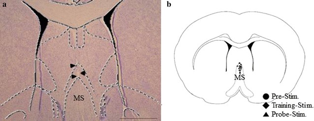
Fig. 3.

Location of electrodes. A representative stained section and an atlas schematic demonstrating the location of electrodes in the medial septum (MS) are shown. a The location of the stimulating electrodes was confirmed using Cresyl violet staining. The arrowheads indicate the tract of the electrode. b The population of electrode locations on an atlas schematic of MS, where circle indicates the location of electrodes in the pre-stimulation group, diamond indicates the location of electrodes in the training-stimulation group, and triangle indicates the location of electrodes in the probe-stimulation group
