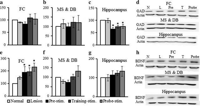
Fig. 6.

Changes in glutamate decarboxylase (GAD) 65/67 and brain-derived neurotrophic factor (BDNF) expression. The expression level of GAD 65/67 was not significantly different in the frontal cortex (FC) (a) or medial septum (MS) and diagonal band (DB) (b) for all the groups compared with that in the normal group. c The hippocampal level of GAD 65/67 was significantly lower relative to the normal group at all stimulation times. d Representative western blotting results. e The expression level of BDNF was significantly higher in all the stimulation groups in the FC. BDNF expression also was slightly higher in the MS and DB (f) and hippocampus (g). h Representative western blotting results. The indices are expressed as a percentage of values for the normal group (mean ± SE, p < 0.05)
