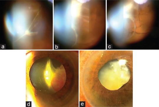
Figure 3.

Slit lamp photographs of the left eye of patient P3. (a) Vitreal and pre-retinal fibrous membrane formation was observed 2 weeks after intravitreal injection of mesenchymal stem cells (MSCs). (b and c) The fibrous tissue severity increased after one month and led to total tractional retinal detachment. (d) Retrolental fibrovascular tissue was visible three months after intravitreal cell injection. (e) Six months after cell injection, mature cataract, ciliary injection, shallow anterior chamber and neovascularization of the iris were present.
