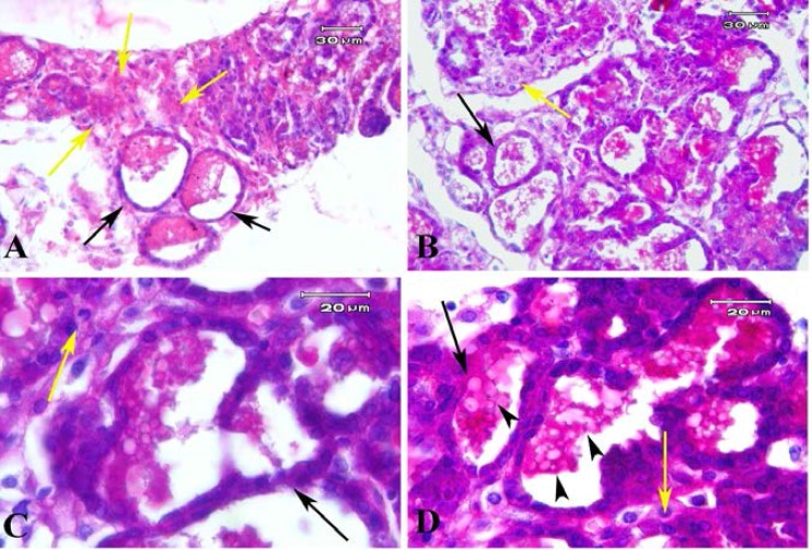
Figure 7.
Photomicrograph of endometriosis lesions in model B stained with PAS method. It demonstrated the endometrial like glands which lined with cuboidal epithelial cells (black arrows) and PAS positive secretion within them (black arrow heads). The stromal cells between gland sections show the PAS positive reaction (yellow arrow

