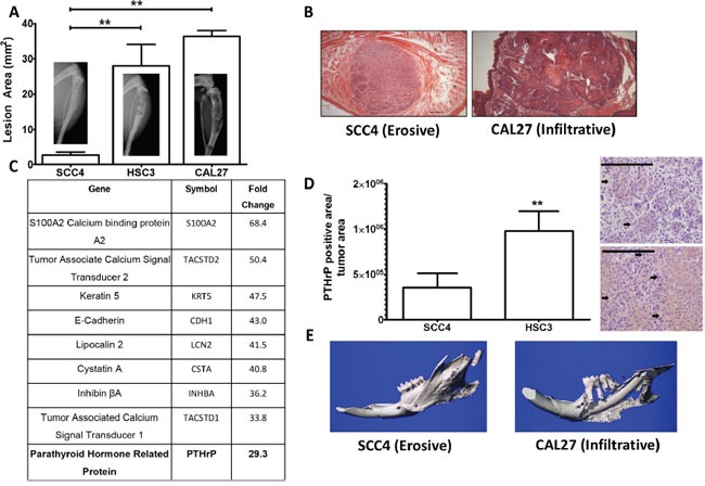
Figure 1. PTHrP mRNA levels predict bony invasion and bone destruction in two mouse models.

A. Tumor model of bone destruction. X-ray data of SCC4 cells injected into the tibiae of athymic mice show significantly less bone destruction than HSC3 and CAL27 cells. B. H&E stained histological sections of SCC4 and CAL27 tumors taken from tip-of-the-tongue injections in athymice mice show morphological differences. SCC4 cells bear an erosive phenotype, while CAL27 cells bear an infiltrative phenotype. C. PTHrP is upregulated almost 30-fold in bony invasive OSCC. The top ten upregulated genes as determined from a genome wide microarray study comparing mRNA levels of CAL27 cells (bony invasive OSCC) vs SCC4 (non-bony OSCC). PTHrP is highlighted because it is known to be essential for OSCC bony invasion. D. PTHrP levels correlate with bony invasion status. SCC4 cells injected into the tibiae of athymic mice show low PTHrP staining by IHC, while HSC3 cells show significantly larger amounts of PTHrP expression, as denoted by the black arrows. (Images at 40X, scale bar is 200μm) E. Orthotopic model of bony invasion. Representative μCT scans of mandibles dissected from mice bearing tumors from masseter muscle injections. SCC4 cells show minimal bone destruction and small amounts of new bone formation, while CAL27 cells show extensive bone destruction.
