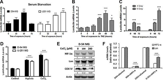
Figure 2. Lon expression is induced by a variety of stressors.

A. The normal culture medium (10% FBS) of D-54 and U-251 cells were replaced by serum-free medium for 3 hours. The cells were then allowed to recover in normal medium for the amount of time indicated. B. D-54 cells were treated with TMZ (500 μM). C. D-54 cells were exposed to 4 or 6 Gy of irradiation. D. D-54 and U-251 cells were cultured in low-oxygen concentrations (1%) or chemically-induced hypoxia (200 μM cobalt chloride) for 24 hours. Cells were collected at the indicated time points and RNA was extracted. qRT-PCR was then performed to measure the Lon mRNA levels. The relative expression levels were normalized by ACTB. E. D-54 cells were treated with 100μM or 200μM of CoCl2 for 24 hours. Western blot was used to detect HIF-1α, LONP1 and COX IV. Actin was used as the internal control. F. Treatment with four different siRNA targeted against the HIF-1α sequence leads to 80-90% decrease of HIF-1α mRNA levels as compared with siControl-treated cells, 72 hours after transfection. Treatment of cells with siRNA targeting HIF-10α reduced Lon mRNA levels (~80% in D-54 and ~50% in U-251), representative data showed. *p < 0.05, **p < 0.01, ***p < 0.001.
