
Figure 4. FOXM1 inhibition enhances radiation induced mitotic catastrophe.
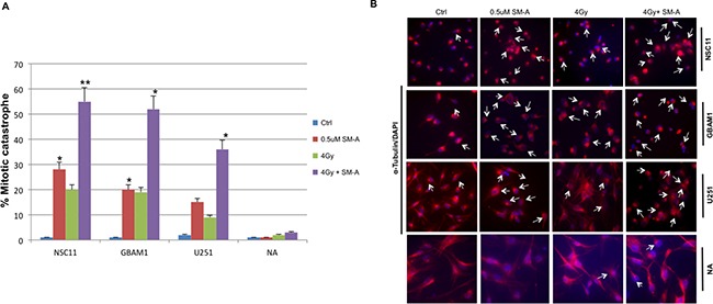
Changes in nuclear morphology of U251, NSC11 and GBAM1 cells following 48hr exposure to Siomycin-A alone or in combination with 4Gy IR. Panel A., quantitation of the percentage of multinucleated cells based on fluorescent microscopy analysis represented as bar graph. Panel B. representative pictures of cells stained with α-Tubulin and DAPI examined under a fluorescent microscope (magnification, 20X). Enlarged cells, containing multiple evenly stained nuclei (multinucleated cells) are characteristic for mitotic catastrophe (arrows). Data presented is the mean ± S.D, from three independent experiments. Student's t test was performed and the level of significance *indicate p < 0.05, **p < 0.005.