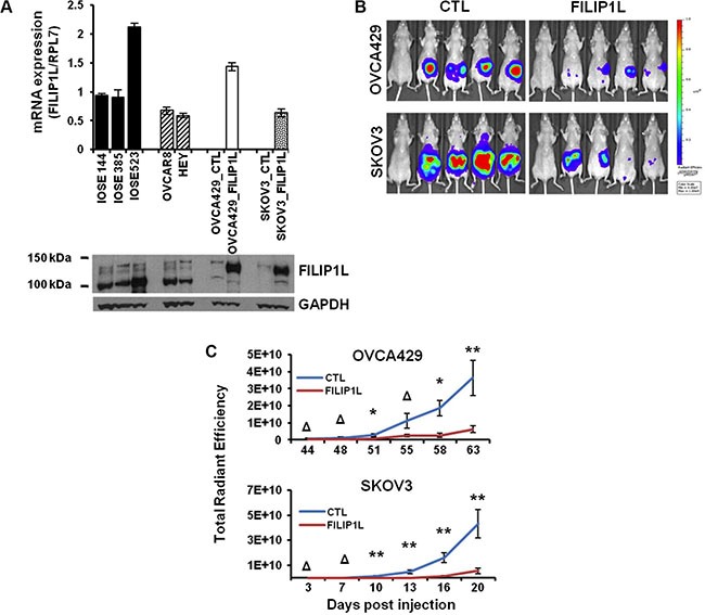
Figure 2. The effect of FILIP1L expression on the metastatic potential of ovarian cancer cell lines in a mouse model.

(A) FILIP1L mRNA levels were determined by qRT-PCR in immortalized normal IOSE cell lines and ovarian cancer cell lines that express high FILIP1L (OVCAR8 and HEY). Also shown are low FILIP1L-expressing OVCA429 and SKOV3 ovarian cancer cells, and their FILIP1L+ derivatives. The y axis represents FILIP1L mRNA expression standardized for the housekeeping gene RPL7. The result is an average of three independent experiments. Immunoblot analysis for FILIP1L in ovarian cells is also shown. Note that the FILIP1L band in FILIP1L+ OVCA429 and SKOV3 cells migrates slower than the other untransfected cells because it is expressed as a fusion protein with GFP. GAPDH blot is shown as the loading control. (B) OVCA429 (106) and SKOV3 (5 × 10⁵) (CTL) and their FILIP1L+ derivatives (FILIP1L) were orthotopically injected into the ovaries of Nude mice. Development of peritoneal metastasis was monitored by IVIS imaging, every 3–4 days for a total of 63 and 20 days for OVCA429 and SKOV3, respectively. Representative images at the time of sacrifice are shown. The first mouse in each series is the negative control. (C) Time course of quantified metastatic tumor growth (8 mice per group). The y axis represents total radiant efficiency ([photons/s]/[μW/cm2]) emitted by iRFP. *,** and Δ indicate P < 0.05, P < 0.01 and NS, respectively. The result is representative of two independent experiments.
