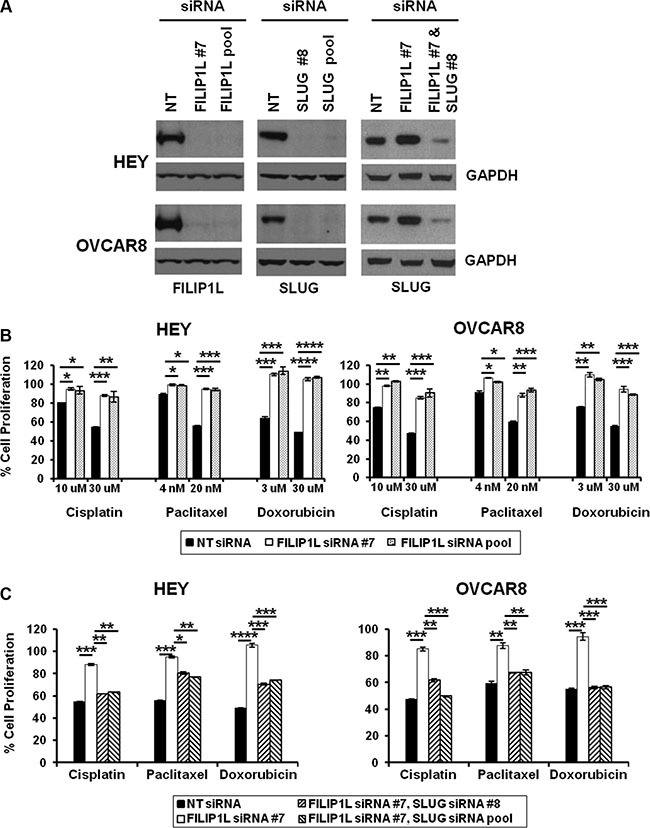
Figure 3. Effects of FILIP1L and an EMT activator on chemoresistance in ovarian cancer cells.

(A) Knockdown of FILIP1L and SLUG was achieved by transient transfection of indicated siRNA. Protein knockdown was shown by immunoblotting. GAPDH blot shown underneath of each blot is the loading control. The result is representative of two independent experiments. (B) Cells were treated with indicated siRNA for 2 days, and with chemotherapy agents of indicated concentration for an additional day. Cell proliferation was measured by WST1 incorporation quantified at OD440-650nm. The y axis represents cell proliferation in the presence of drug as a percentage of untreated control. (C) The same experimental procedures were used as described in section B with indicated siRNA treatment but only 30 μM cisplatin, 20 nM paclitaxel or 30 μM doxorubicin were used. *, **, *** and **** indicate P < 0.05, P < 0.01, P < 0.001 and P < 0.0001, respectively. The result is an average of three independent experiments.
