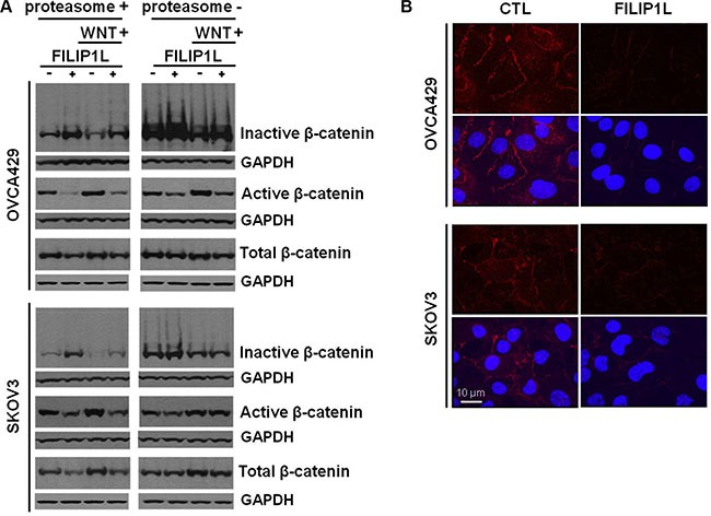
Figure 4. Effect of FILIP1L on β-catenin degradation by proteasomes.

(A) Parental and FILIP1L+ OVCA429 and SKOV3 clones were treated with solvent or 40 mM LiCl for 24 h (WNT+). Cell lysates were immunoblotted with antibodies against inactive phospho-β-catenin, active non-phospho-β-catenin, and total β-catenin. The experiment was also performed in the presence of 5 μM MG132 (proteasome-). GAPDH blot is shown as the loading control. (B) FILIP1L reduces active β-catenin. OVCA429 and SKOV3 clones were immunofluorescently stained for active β-catenin (red) and nuclei counterstained with DAPI (blue). Red only and merged images are shown. The result is representative of three independent experiments.
