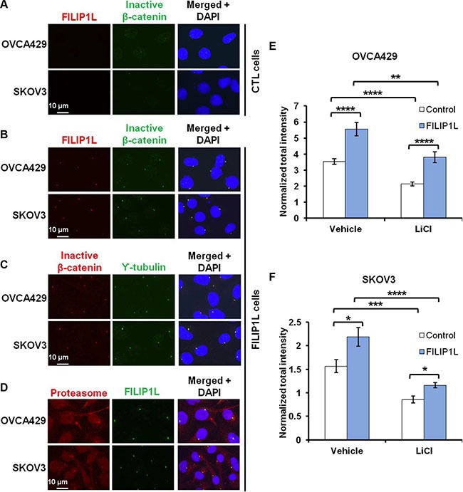
Figure 5. FILIP1L increases inactive β-catenin at centrosomes.

(A–B) OVCA429 and SKOV3 clones were immunofluorescently stained for FILIP1L (red) and inactive β-catenin (green). Staining is shown in either control (A) or FILIP1L-expressing (B) clones. (C–D) FILIP1L-expressing clones were immunofluorescently stained for inactive β-catenin (red) and γ-tubulin (green) (C) and for proteasome 19S-S7 subunit (red) and FILIP1L (green) (D). Nuclei were counterstained with DAPI (blue). Merged images are also shown. (E–F) The quantified data from 200-250 cells is also shown. *, **, *** and **** indicate P < 0.05, P < 0.01, P < 0.001 and P < 0.0001, respectively. The result is an average of three independent experiments.
