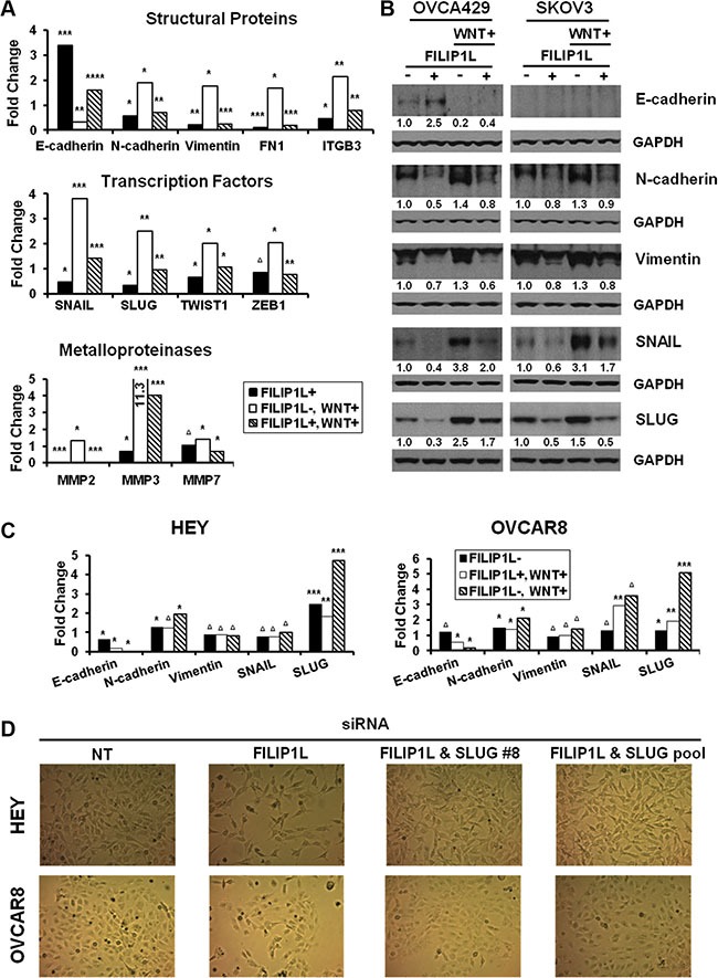
Figure 6. Regulation of EMT markers by WNT signaling and FILIP1L.

(A) Parental or FILIP1L+ OVCA429 clones were treated with solvent or 40 mM LiCl for 24 h (WNT+). mRNA levels of EMT markers were measured by qRT-PCR. The y axis represents fold change over the control (solvent-treated parental cells). Each value was also standardized for the housekeeping gene GAPDH. The result is an average of three independent experiments. (B) Immunoblot analysis for the indicated EMT markers from OVCA429 and SKOV3 clones treated with the same methods as in section A. GAPDH blot is shown as the loading control. Values indicate the quantified each protein amount normalized to the loading control GAPDH. Note that endogenous E-cadherin protein levels could not be detected due to low levels of expression in SKOV3 cells. The result is representative of three independent experiments. (C) HEY and OVCAR8 cells were treated with either non-targeting or FILIP1L siRNA, and with solvent or 40 mM LiCl for 24 h (WNT+). mRNA levels of EMT markers were measured as described in section A. The result is an average of three independent experiments. (D) HEY and OVCAR8 cells were treated with indicated siRNA for 2 days and cell morphology was imaged with light microscope. *, **, ***, **** and Δ indicate P < 0.05, P < 0.01, P < 0.001, P < 0.0001 and NS, respectively.
