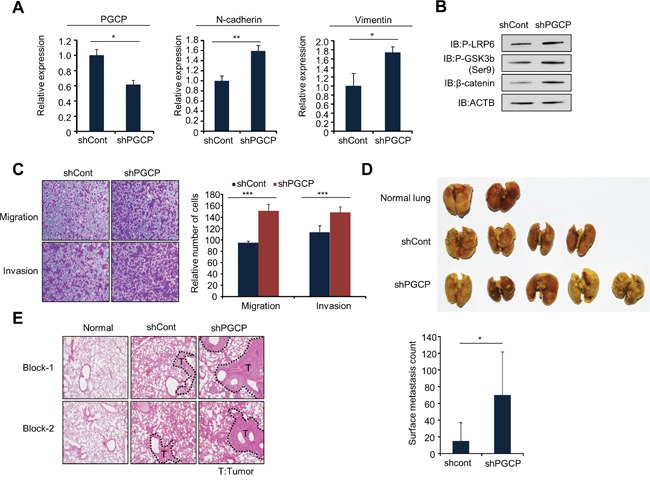
Figure 5. Knockdown of PGCP promotes lung metastasis in vivo.

A. Quantitative RT-PCR analysis of PGCP, N-cadherin and vimentin cDNA was isolated from shCont and shPGCP stable cell lines. The p values were calculated using Student's t-test (*p<0.05, **p<0.01). B. Western blot analysis of phospho-LRP6, phospho-GSK3β (Ser9) and β-catenin. ACTB was used as an internal control. C. Migration and invasion assay with shCont and shPGCP cells. Migrating and invasive cells were fixed with methanol after 24 h stained with crystal violet. The p values were calculated using Student's t-test ***p <0.001. D. Gross lung metastases. shCont and shPGCP cells (2 × 10⁶) were injected into BALB/c nude mice through tail vein, and lung metastases were assessed 4 weeks after injection (n=5 per group) (top). Quantification of metastatic nodules (bottom). p values were calculated using Student's t-test *p <0.05. E. Representative images of hematoxylin and eosin staining of lung metastases. T, tumor.
