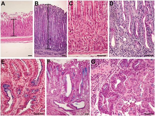
Figure 1. Development of histopathological lesions of the gastric mucosa after Helicobacter pylori infection in iqgap1+/- mice.

Representative images of histopathologic features of the gastric mucosa of uninfected (A, C) or H. pylori SS1-infected (B, D, E, F, G) iqgap1+/- mice after 12 months. (A, C) Normal gastric mucosa. (B) H pylori-induced hyperplasia compared to the normal thickness of the mucosa of an uninfected control mouse (A). (D) Mucinous metaplasia with mucins producing cells replacing parietal and chief cells observed in normal gastric mucosa of a control mouse (C). (E) Pseudo-intestinal metaplasia with cells resembling enterocytes and expressing mucins mainly at the apical surface. (F-G) Dysplasia with gastrointestinal intraepithelial neoplasia (GIN) penetrating the muscularis mucosa. (G) GIN with an area of less cohesive dysplastic cells. Images are representative of the lesions obtained with Helicobacter felis and H. pylori SS1 and HPARE strains in WT and iqgap1+/- mice. Scale bar = 50 μm.
