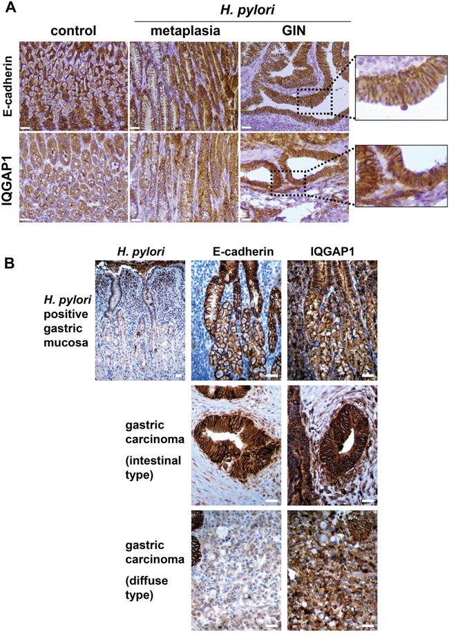
Figure 3. Expression of IQGAP1 and E-cadherin in gastric mucosa of mice and humans infected by Helicobacter pylori and in GIN and gastric carcinoma.

Representative images of immunohistochemistry detection (in brown) of H. pylori, IQGAP1, and E-cadherin on gastric PET-sections. (A) Gastric tissue sections from uninfected (control) or H. pylori SS1-infected wild type (WT) mice (12 months) having developed metaplasia of the mucinous type (left side of the metaplasia images) and/or pseudo-intestinal type (right side of the metaplasia images) and GIN. (B) Representative gastric tissue sections from only gastric cancer patients with H. pylori associated chronic gastritis (n = 6) (top row), with moderately differentiated intestinal-type adenocarcinoma (n = 6) (second row) and with diffuse type adenocarcinoma comprised of E-cadherin-negative isolated signet ring cells (n = 3) (third row). Scale bars, 25 μm.
