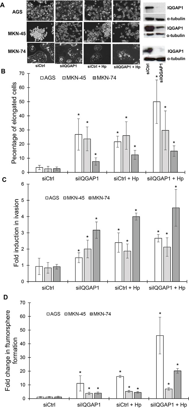
Figure 4. IQGAP1 inhibition mimicks Helicobacter pylori induced effects on gastric epithelial cell morphology, invasion capacities and tumorsphere formation.

AGS, MKN-45 and MKN-74 cells were transfected in a double round of transfection with the negative control (siCtrl) or IQGAP1 (siIQGAP1) siRNA, and infected or not with H. pylori 7.13 for 18 to 24 h. (A) Representative phase-contrast images of transfected cells after infection or not with H. pylori 7.13; and representative images of western blotting experiments showing the inhibition of expression of IQGAP1 by siIQGAP1 compared to siCtrl transfected cells. α-tubulin detection was used as a loading control of equal amounts of proteins. Scale bar: 10 μm. (B) Quantification of the percentage of cells harboring an elongated phenotype. (C) Quantification of cellular invasion after 18 h of infection in the Transwell invasion assay. * p<0.05 vs. uninfected siCtrl cells. (D) Quantification of spheroids obtained under non-adherent culture conditions after 5 days of infection in the tumorsphere assay.
