
Figure 1.
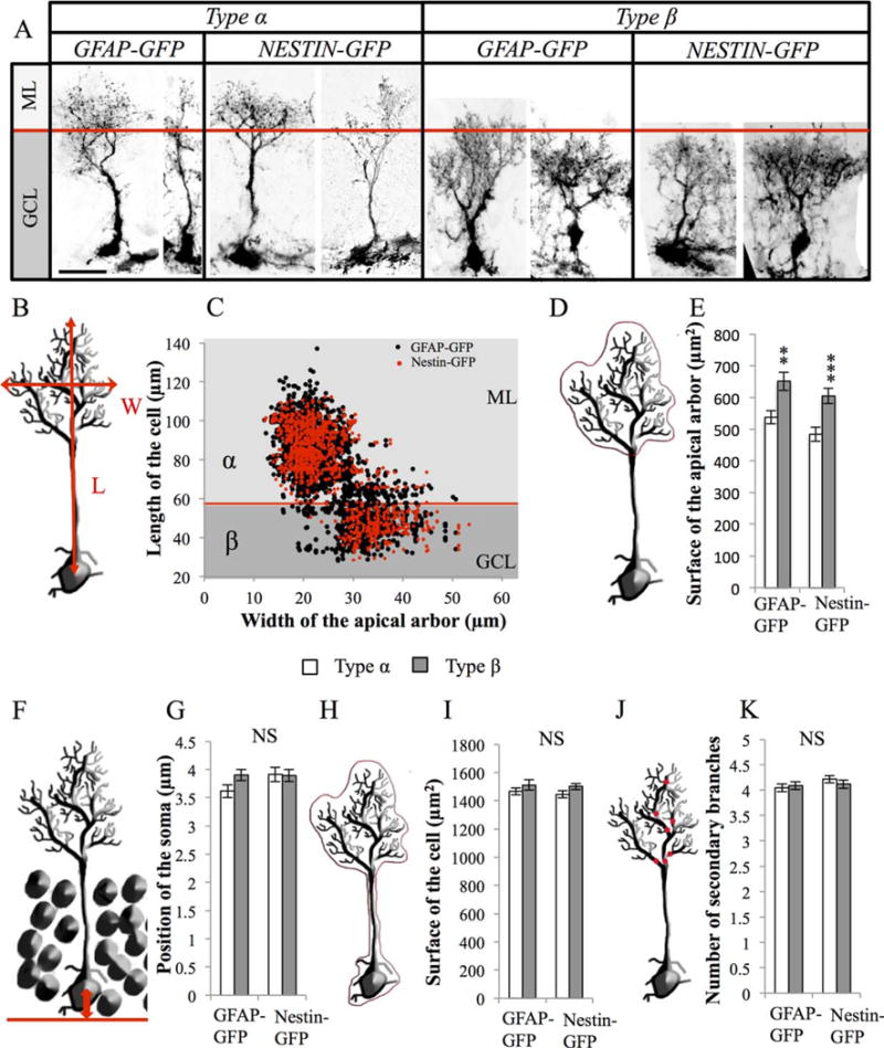
Morphometrical parameters of radial glia-like (RGL) cells. (A): Confocal maximal projection micrographs of types α and β RGL cells in glial fibrillary acidic protein (GFAP)-green fluorescent protein (GFP) and Nestin-GFP mice. (B): Drawing of a RGL neural stem cell illustrating the measurements of length and width of the cell. (C): Scatter graph of all RGL cells analyzed morphometrically (n = 2472 for GFAP-GFP mice and n = 1150 for Nestin-GFP mice). (D, E): Schematic illustration (D) and histogram (E) of the projected surface of the dendritic arbor of secondary processes in types α and β cells. (F, G): Schematic illustration (F) and histogram (G) of the position of the soma of type α and the type β cells relative to the hilar border of the granule cell layer. (H, I): Schematic illustration (H) and histogram (I) of the total surface of types α and β cells. (J, K): Drawing (J) and histogram (K) of the number of branches of the main process of types α and β cells. Scale bar: 20 μm. Bilateral Student’s t test **, p < 0.01; ***, p < 0.001. Each value represents the mean ± SEM. Abbreviations: GCL, granule cell layer; GFAP, glial fibrillary acidic protein; GFP, green fluorescent protein; ML, molecular layer.