
Figure 5.
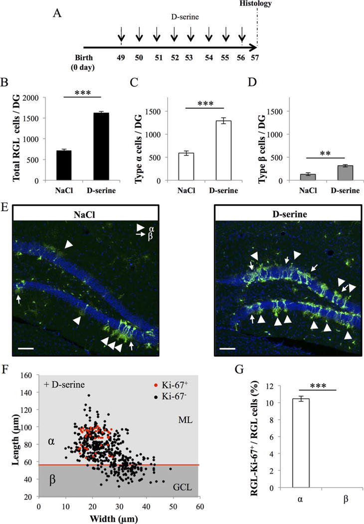
Number of types α and β cells are increased by D-serine injection. (A): Experimental timeline: Nestin-GFP mice were injected with D-serine, daily for 8 days, starting at 49 days after birth and were killed 1 day after the last D-serine injection. (B): Histogram of the total number of radial glia-like (RGL) cells in the dentate gyrus (bilateral Student’s t test p < 0.001). (C): Histogram of the number of type α cells in the dentate gyrus (bilateral Student’s t test p < 0.001). (D): Histogram of the number of the type β cells in the dentate gyrus (bilateral Student’s t test p < 0.01). (E): Confocal maximal projection micrographs of hippocampal sections. (F): Scatter graph of RGL cells dimensions. Red dots: Ki-67+ cells N = 45, black dots Ki-67− cells N = 500. (G): Histogram showing the percentage of types α and β cells expressing Ki-67. Bilateral Student’s t test. Scale bars = 100 μm. N = 4 animals per group. Each value represents the mean- ± SEM. **, p < 0.01; ***, p < 0.001. Abbreviations: DG, Dentate Gyrus; GCL, granule cell layer; ML, molecular layer; RGL, radial glia-like.