Fig. 2.
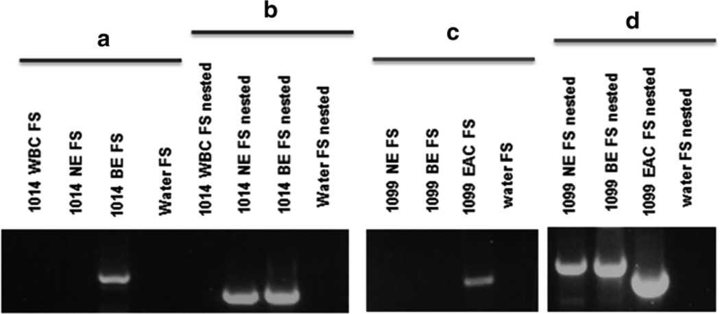
Validation of results. (a) Diagram of the PCR validation scheme for putative insertions, the 3′ end of the LINE-1 insertion is pictured adjacent to a poly A tail. The nested empty site and filled site primers are flanking the empty and filled site primers. In a nested PCR, the nested primers are used in the first of two reactions. One and a half µL of product from the first reaction (with ES and FS primers) are used as template in a second PCR with the nested primers to amplify difficult or rare products. (b) Two examples of validations for insertions present in only tumor and absent from normal DNA. On the left, a PCR result depicting both the empty site (ES) and filled site (FS) products for both the normal and tumor DNA samples from patients. Only in the tumor of patient 11 is a filled site band present confirming the insertion is present. In the image on the right side on b, a PCR depicting another validation of a somatic insertion present in BE and absent from normal esophageal and white blood cell DNA. There is only a band present in the BE sample for the FS PCR; however, the ES PCR has bands for all three DNA samples as a positive control. (c) An insertion sequence with the unique genomic DNA (blue), target site duplications (purple), LINE-1 sequence (red), and the poly-A tail sequence (orange) [15]

