Figure 2.
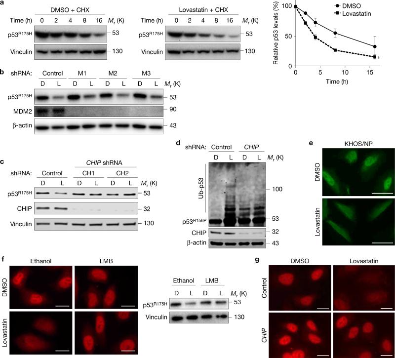
Statins induce mutp53 degradation by CHIP. (a) WB for p53 and vinculin using SK-Br-3 cells treated with cycloheximide (CHX, 50 nM) for the indicated time periods (h) following pre-incubation with DMSO or lovastatin (4 μM) for 12 h. Right: graph showing relative p53R175H levels compared with those without CHX. Error bars, means ± s.d. (n = 3 independent experiments), *P < 0.05; Student's t-test (two-tailed). (b) WB for the indicated proteins using SK-Br-3 cells infected with non-silencing control or MDM2 (M1, 2, 3) shRNA-encoding lentiviral vectors and treated with DMSO (D) or lovastatin (L) at 4 μM for 24 h. (c) WB for the indicated proteins using SK-Br-3 cells infected with lentiviral vectors encoding non-silencing control or CHIP (CH1, 2) shRNAs and treated with D or L (4 μM) for 24 h. (d) Ubiquitin assays for p53R156P and WB for the indicated proteins. KHOS/NP cells with or without CHIP knockdown were transfected with an ubiquitin-encoding plasmid, treated with D or L (4 μM) for 22 h, further incubated with 30 μM of MG132 for 6 h, and harvested with hot SDS lysis buffer. (e) Immunofluorescence for p53R175H in KHOS/NP cells treated with D or L for 18 h. Scale bars, 50 μm. (f) Immunofluorescence (left) and WB (right) for p53R175H using SK-Br-3 cells treated with D or L along with vehicle (ethanol) or leptomycin B (LMB, 50 nM) for 18 h. Scale bars, 50 μm. (g) Immunofluorescence for p53R175H using SK-Br-3 cells infected with control or CHIP shRNA-encoding lentiviral vectors and treated with DMSO or lovastatin. Scale bars, 50 μm. Statistics source data for a are provided in Supplementary Table 1. Additional results are shown in Supplementary Fig. 2. Unprocessed original scans of blots are shown in Supplementary Fig. 8.

