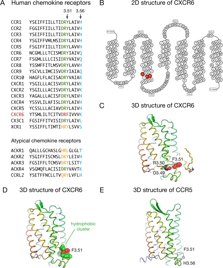
Fig 1. Sequences and structural features of CXCR6 around the DRF motif.
A: Sequence of TM3 (residues 3.37–3.56) in human chemokine receptors. The DRY motif (residues 3.49–3.51) is highlighted in green, the DRF motif of CXCR6 in red, other motifs at this position in orange, and residues at position 3.56 in blue. B: 2D structure of the human CXCR6 (obtained from the GPCRdb [67]), with DRF motif highlighted in red. C/D: Snapshot from the molecular dynamics simulation of the 3D model of the human CXCR6. The residues in the DRF motif (D1263.49, R1273.50 and F1283.51) are shown as spheres, and a lipid molecule in contact with F1283.51 is shown as sticks (C). The cluster of hydrophobic residues around F1283.51 is shown in D. The rest of protein side chains, lipids, water molecules, and ions are not displayed for clarity. E: 3D structure of the human CCR5 receptor, showing the hydrogen bond between residues at positions 3.51 and 3.56.

