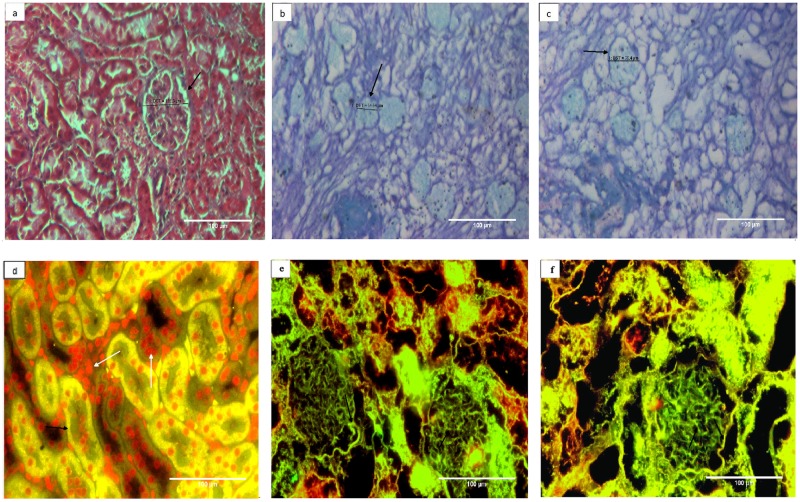
Fig 3. Collagen content of basement membrane is well retained whether or not the kidneys were cryostored prior to decellularization.
Both Masson’s trichrome staining (photomicrogrpahs a-c) and immunohistochemistry using anti collagen IV antisera (photomicrograph d-f) were used to analyze the effect of cryostorage on collagen status of the decellularized structures. Masson’s trichrome stains collagen blue, cellular component red and nuclei black. The blue stained structures (black arrows) showed well retained collagen component of the ECM from a) represented control kidney from group I, b) decellularized kidney from group II, c) decellularized kidney from group III. Almost similar extent of staining patterns among group II and III demonstrated that decellularization process of kidneys did not affect the collagen content whether the tissues were cryopreserved or not. Absence of red and black stain re-confirms the absence of any cellular material and nucleus in these decellularized kidneys. With respect to immunohistochemistry (fig d-f) green fluorescence represented collagen IV and the red fluorescence indicated cell nucleus (Propidium iodide, PI stain). Photomicrographs represented d) control kidney from group I, e) decellularized structures from freshly isolated kidney, group II and f) decellularized structure from cryostored kidney, group III at 200X. Where-ever shown, the white arrows represent renal cells (red fluorescence) and black arrows represent collagen 1V (green fluorescence). It is clear from the Figs that after decellularization overall presence of ECM component collagen IV in glomerulus and kidney structure (black arrow) is preserved and cells are completely removed as no red fluorescence observed when compared with control.

