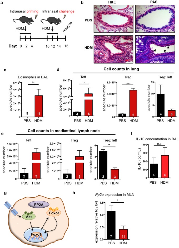
Fig 1. HDM potently induces allergic airway inflammation despite significant T regulatory cell recruitment.
(a) Allergic airway inflammation was induced with three separate priming and challenge doses of house dust mite (HDM) over 14 days. Disease severity was assessed at day 15. (b) Histological assessment of inflammation in lung sections of representative HDM-treated and PBS-treated mice. * in H&E images indicates inflammatory infiltrate and arrowheads in PAS images indicate mucus. (c) Eosinophils were quantified in the bronchoalveolar lavage (BAL) by flow cytometry (CD45+Cd11b+SiglecF+CD11c−). Treg (CD4+CD25+FoxP3+) Teff (CD4+CD25+FoxP3−) populations in the lung parenchyma (d) and mediastinal lymph nodes (MLN) (e) were quantified by flow cytometry. (f) IL-10 concentration in BAL was quantified by ELISA. (g) Schematic of PP2A-FoxO1 axis. (h) PP2Acα sub-unit transcript levels were measured by RT-PCR. Transcript levels in HDM-treated mice were normalized to PBS-treated controls. Numbers in the bars of c, d, e, f and h represent the number of animals analyzed. Significance in all panels was determined by two-tailed students’ T-test: *<0.05; **<0.01; ****<0.0001.

