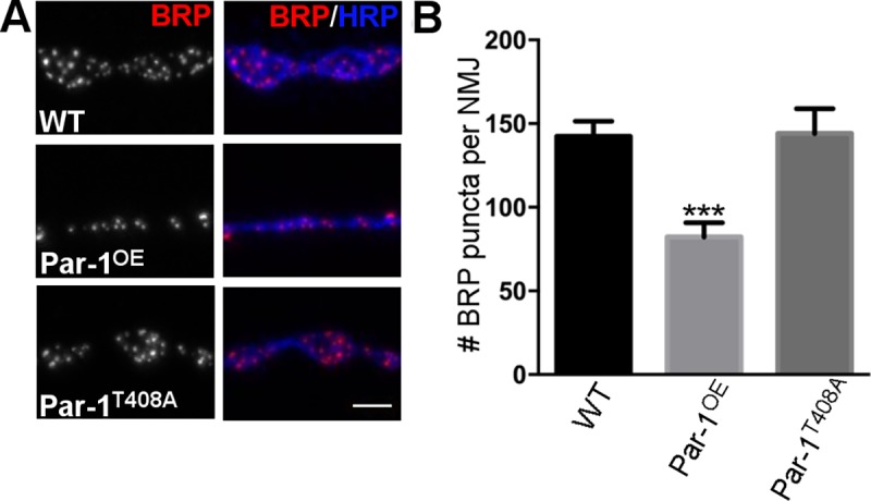
Fig 3.

A) Representative images from WT, Par-1OE and Par-1T408A showing the NMJ synapses labeled with anti-BRP (Red) and anti-HRP (Blue) antibodies B) Quantification of BRP puncta per NMJ. N = 10, *** = p≤0.0001. Scale bar = 10μm. Error bars represent S.E.M.
