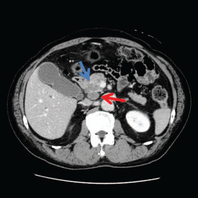
Figure 1.

Contrast-enhanced computed tomography images showing a tumor in the pancreatic head measuring 3.0 × 2.5 cm in diameter and invading the common bile duct (indicated by a blue arrow). Lymphadenopathy was observed posterior to the pancreatic head (red arrow).
