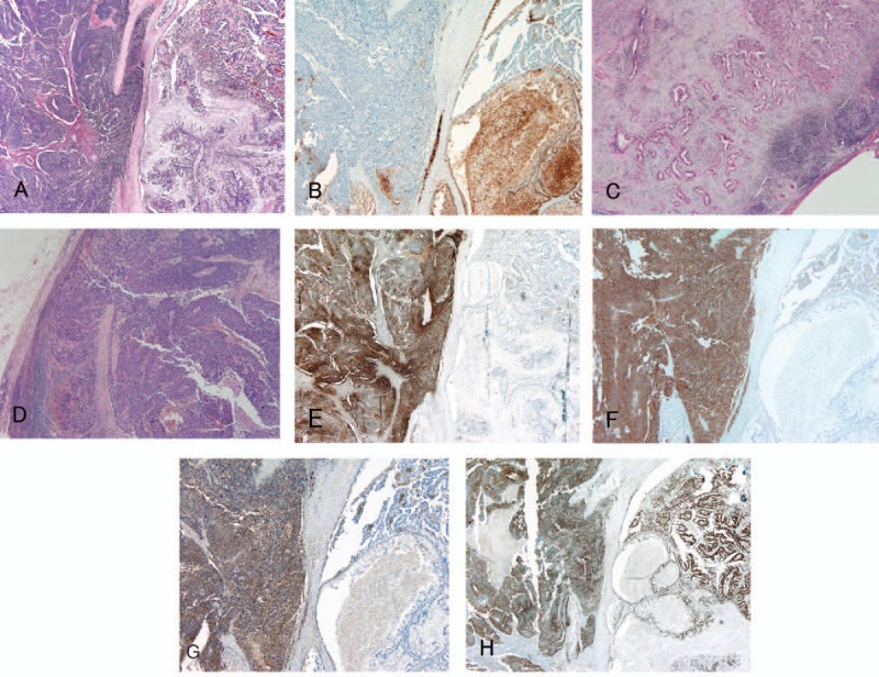
Figure 2.

Microscopic and immunohistochemical appearance of the tumor. (A) Primary tumor (hematoxylin and eosin staining, original magnification ×20). The primary lesion was composed of a well-differentiated adenocarcinoma component and a poorly differentiated NEC component, each tightly intermingled. (B) Immunohistochemical staining of the primary lesion for CEA (original magnification ×20). CEA was limited to the well-differentiated adenocarcinoma component of the primary lesion. (C) LN no. 12 exclusively contained the well-differentiated adenocarcinoma component (original magnification ×20). (D) LN no. 13 exclusively contained the poorly differentiated NEC component (original magnification ×20). (E) Immunohistochemical staining of the primary lesion for chromogranin A (original magnification ×20). Chromogranin was limited to the poorly differentiated NEC component of the primary lesion. (F) Immunohistochemical staining of the primary lesion for synaptophysin (original magnification ×20). Synaptophysin was limited to the poorly differentiated NEC component of the primary lesion. (G) Immunohistochemical staining of the primary lesion for NSE (original magnification ×20). NSE was limited to the poorly differentiated NEC component of the primary lesion. (H) Immunohistochemical staining of the primary lesion for p53 (original magnification ×20). p53 was ubiquitously expressed in both the well-differentiated adenocarcinoma and poorly differentiated NEC components of the primary lesion. The well-differentiated adenocarcinoma component can be seen on the right sides and the poorly differentiated NEC component can be seen on the left (E, F, G, and H). CEA = carcinoembryonic antigen, LN = lymph node, NEC = neuroendocrine carcinoma, NSE = neuron-specific enolase.
