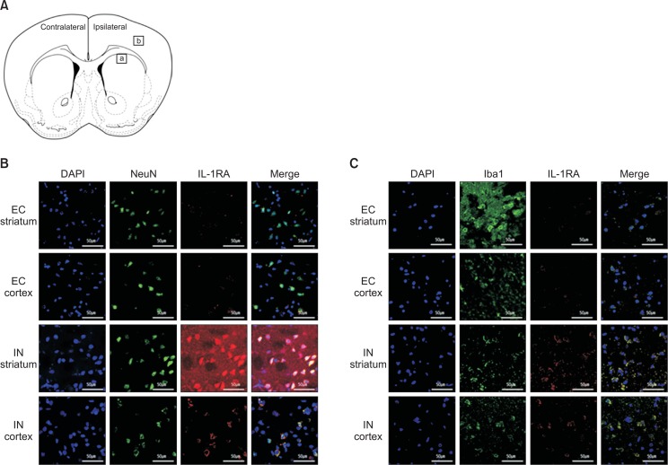
Fig. 4.
Distribution of human IL-1RA in the striatum and cortex at 6 h after intranasal administration. A coronal section of the rat brain (A). The square areas represent the location where the following immunostaining images were taken (a, striatum and b, cortex). After intranasal administration, human IL-RA was detected with strong immunoreactivity and largely co-localized with NeuN (B) and Iba-1 (C) in the striatum and cortex of the rats in the IN group. On the other hand, the immunoreactivity in the EC group is minimal.

