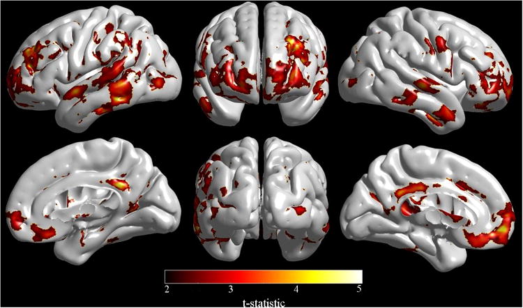
Figure 3.

Cortical modeling showed significant cortical thickness reductions in HIV-infected patients compared to HIV-uninfected participants specifically within the right primary motor and sensory cortex, lateral temporal and frontal lobes, and posterior cingulate, orbitofrontal cortex and left anterior cingulate. (Image created with SurfStat http://www.math.mcgill.ca/keith/surfstat/).
