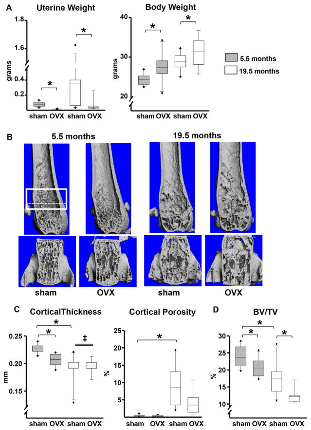
Fig. 1.
The effects of aging on cortical bone are independent of estrogen status. Mice were either sham-operated (n =10) or ovariectomized (5.5 months, n =10; 19.5 months, n =9) for 6 weeks. (A) Uterine and body weights. (B) Representative micro-CT images of femurs (top) and L5 vertebrae (bottom). The box depicts a region of 151 consecutive slices (12 μm/slice) analyzed for cancellous bone volume, as detailed in Supplemental Methods. The most proximate 90 slices, contained between the dotted line and the upper box line, were used for the measurements of cortical porosity to completely avoid the growth plate. (C) Femoral cortical (Ct.) thickness by micro-CT in the midshaft region and cortical porosity in the distal metaphysis. (D) Cancellous bone (bone volume/tissue volume [BV/TV]) in the 5th lumbar vertebra. Boxes depict values from the 25th to 75th quartiles, the middle line depicts the mean, and the vertical whiskers show the 10th and 90th percentiles; values outside this range are plotted as dots. Statistical significance calculated by two-way ANOVA, *p <0.05; age × surgery interactions denoted as ‡.

