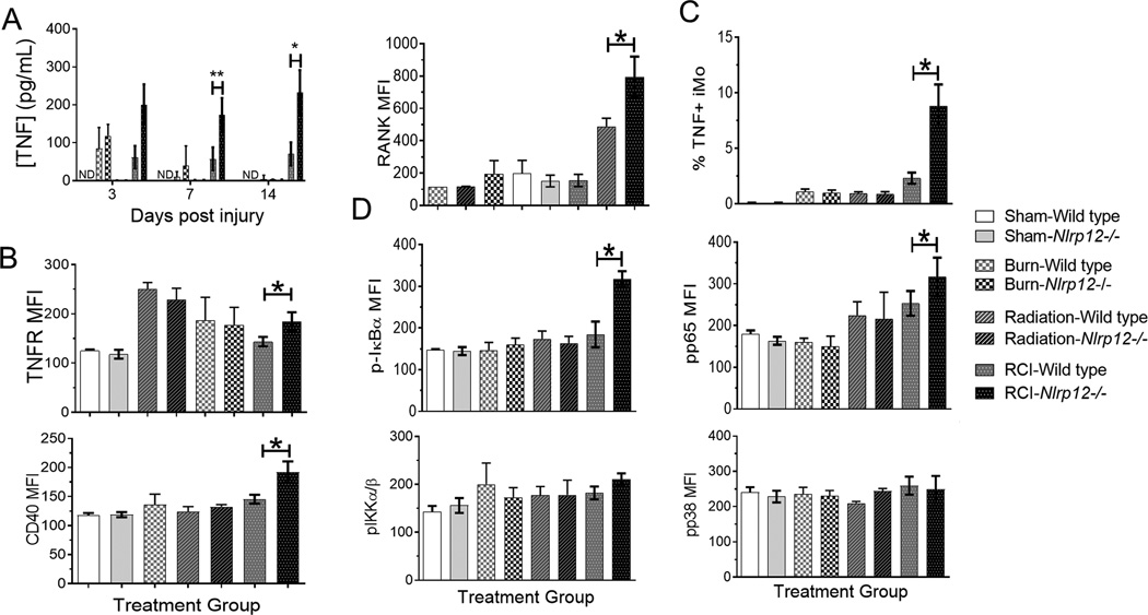
Figure 4. Nlrp12−/− animals have increased serum cytokine and bone marrow receptor expression following combined injury.
Wildtype C57BL/6 or Nlrp12−/− mice were subjected to sham or combined radiation and burn injury (RCI). The concentration of (A) TNF was quantified using ELISA in serum 3, 7, and 14 days post injury. We also analyzed mean fluorescent intensity of (B) TNFR, CD40, and (C) RANK on bone marrow cells harvested at 14 days post injury using flow cytometry. (D) The percentage of TNF producing iMos was determined using intracellular staining and flow cytometry. (E) The level of phopso-IκBα, phospo-IKKα/β, phosphor-p65, and phospho-p38 was quantified using intracellular staining and flow cytometry. Data represented as mean ± SEM, with statistical significance defined as ** p<0.005 by Student’s t test with n=5 mice per group, with experiments performed in triplicate.

