
Figure 4. Exposure to the SDR stressor increased the percentage of IL-1β producing myeloid cells.
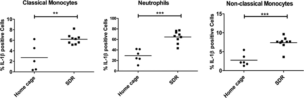
Spleens were harvested from SDR stressed mice and home cage control mice and single cell suspensions analyzed by RNA flow cytometry using the gating strategy outlined in supplemental Fig. 1 and a RNA probe set specific for IL-1β mRNA. The percentage of IL-1β mRNA positive cells was determined for individual home cage and SDR mice. Statistical analysis was performed by Student t test with Welch’s correction for unequal variance. N=5–9 mice per group ** p<0.01, *** p<0.001