
Figure 5.
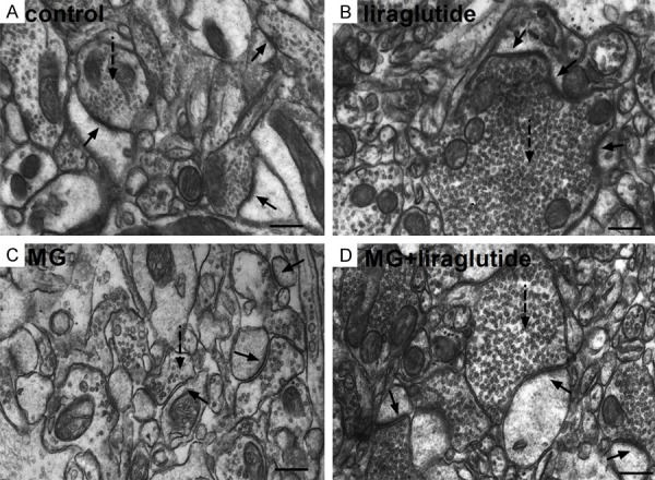
Liraglutide alleviates MG-induced synaptic structure degradation (TEM). (A, B) Chemical synapses in mice that received NS (A) or liraglutide (B) appeared normal: the postsynaptic density was centralized on the intracellular surface of the opposing postsynaptic membrane; synaptic vesicles were observed inside the presynaptic membrane; the synaptic cleft was narrow and with parallel membranes either side. (C) In mice that received MG, synaptic structure was destroyed, with fewer synaptic vesicles, a smaller postsynaptic region, deformed anterior region, and a smaller postsynaptic density; the synaptic cleft was markedly widened and the postsynaptic membrane was swollen. (D) In the MG + liraglutide group, vesicles were evenly distributed in the presynaptic area, and the postsynaptic membrane was slightly thickened. Compared with the MG group, the synaptic form was more regular and the cleft was recognizable. Solid arrow, synapse; dotted arrowheads, synaptic vesicles. n = 3 mice per group. Scale bar = 300 nm. TEM, transmission electron microscopy; MG, methylglyoxal.