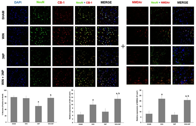
Figure 7.
Effect of WIN55,212-2 and/or 3-NP on the immunofluorescent co-localization of NMDAr and CB1 receptor in neuronal cells (NeuN) from the rat striatum. Animals received WIN55,212-2 (1 mg/kg, i.p. once a day × 6 days prior to 3-NP) or vehicle, plus 3-NP (500 nmol/µL, intrastriatal bilateral dose) and striata were histologically processed and stained with fluorescent antibodies against the mentioned proteins. In the upper panel, micrographs of striatal tissue showing the localization of NeuN (green), CB1 (red in the third, fourth and fifth columns), and NMDAr (red in the sixth, seventh and eight columns) from rats of all treatments are shown at a 20× magnification. In the bottom, counting of neuronal cells and fluorescence intensities is depicted. Mean values ± S.E.M of n=5 rats per group are shown; aP<0.05, different of Sham; bP<0.05, different of 3-NP group. Two-way ANOVA followed by Bonferroni’s test.

