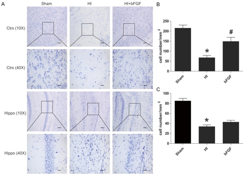
Figure 2.

bFGF inhibits neuronal cell death after HI brain injury. Coronal brain sections were obtained from the sham group, HI group and bFGF-treated group at 1 week after HI. A. Nissl staining pictures of the cortex and hippocampus of the lesioned (ipsilateral) side are shown. B. Analysis of neuron numbers in the boxes of the cortex in different groups. *represents P<0.01 versus the sham group, and #represents P<0.05 versus the HI group. C. Analysis of the neuron numbers in the sections of the hippocampus in different groups. *represents P<0.01 versus the sham group. All data represent the mean value ± SEM, n=4.
