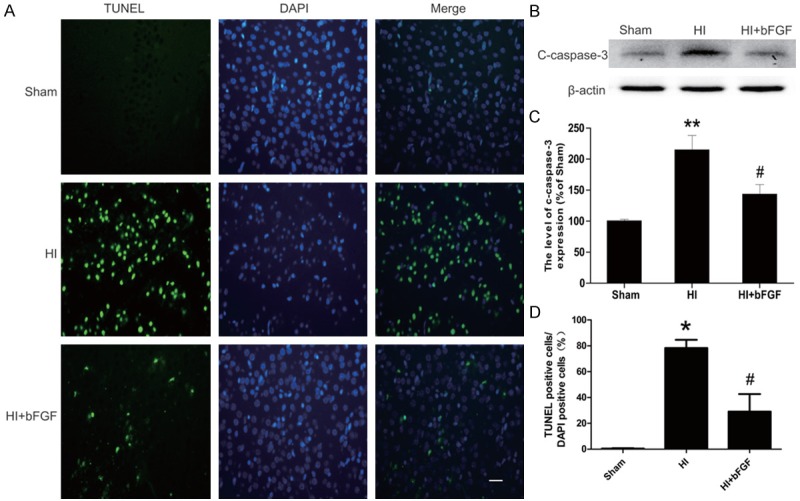
Figure 3.

bFGF attenuates neuronal apoptosis and the caspase cascade in the injury areas of rats after HI. A. TUNEL immunofluorescence (green) and DAPI (blue) staining of sections from the cortical area of the lesioned side in each group. B. Protein expression levels of cleaved caspase3 in the brains of the sham rats, HI rats and HI rats treated with bFGF. C. Optical density analysis of cleaved caspase3 protein. *represents P<0.01 versus the sham group, and #represents P<0.05 versus the HI group. D. Quantitative analysis of TUNEL staining data from A. The percentage of TUNEL-positive cells was expressed as the number of TUNEL-stained nuclei divided by the total number of DAPI-stained nuclei. *represents P<0.01 versus the sham group, and #represents P<0.05 versus the HI group. All data represent the mean value ± SEM, n=4.
