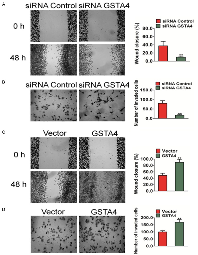
Figure 3.

The effects of GSTA4 on migration and invasion of HCC cells in vitro. A. A wound healing assay was used to evaluate the migration of MHCC97 cells after silencing GSTA4. 24 hours after the transfection of siRNA, cells were wounded and monitored with a microscope every 48 hours. The migration was determined by the rate of cells filling the scratched area. B. Cell invasion was assayed in Transwell coated with Matrigel. MHCC97 cells crossed the Matrigel-coated filter were fixed, stained and counted. Representative pictures of the bottom surface were shown. Five random microscopic fields were counted for each group. The results presented were an average of five random microscopic fields from three independent experiments. The data are presented as mean ± SD. For indicated comparison, **P < 0.01 vs. control cells. C. Representative pictures of (left panel) and quantification (right panel) of migration in HepG2 cells transfected with GSTA4 were analyzed using a wound healing assay. 24 hours after the transfection of GSTA4 plasmid, cells were wounded and monitored with a microscope every 48 hours. The migration was determined by the rate of cells filling the scratched area. D. Significant induction of invasion was observed after transfected over-expression GSTA4 in HepG2 cells. The data were presented as mean ± SD. For indicated comparisons, **P < 0.01 vs. control cells.
