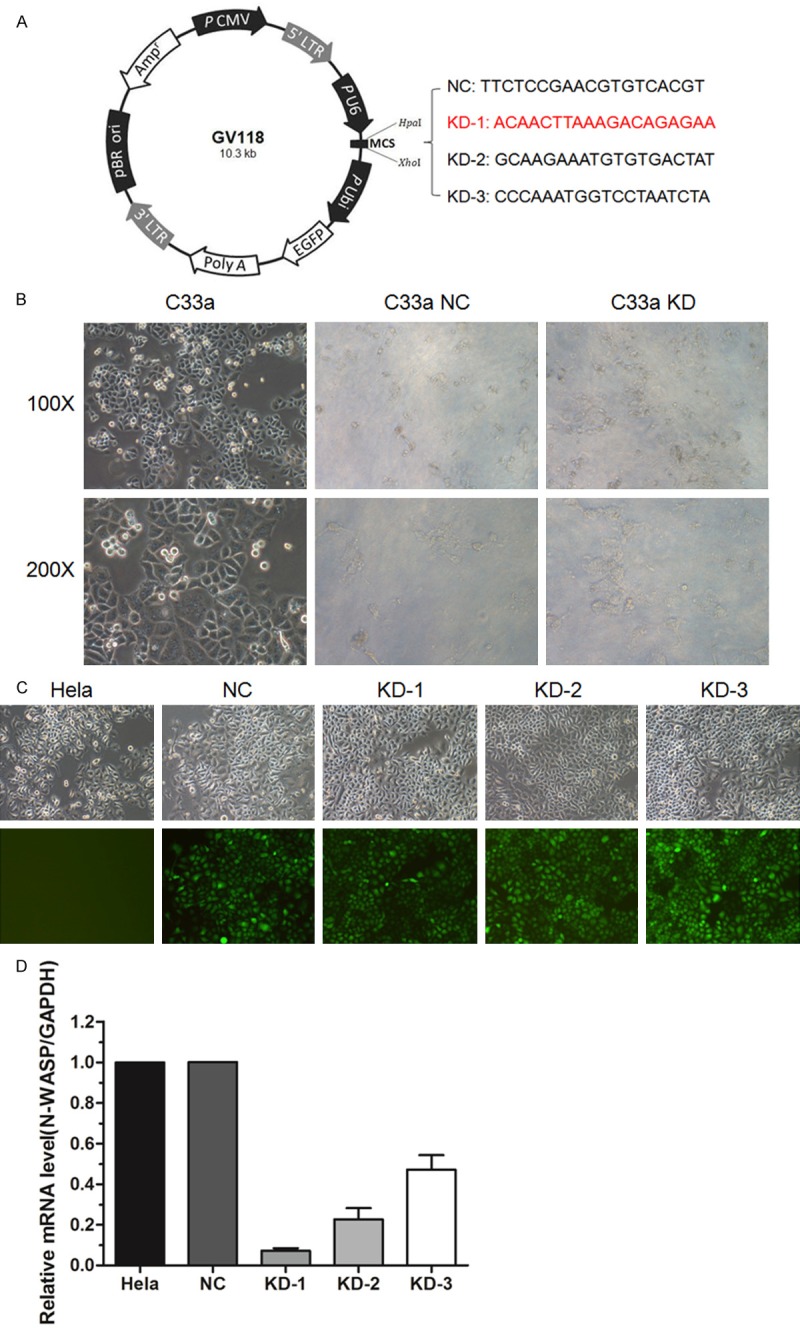
Figure 3.

Establishment of N-WASP silenced cervical cell lines. A. Design of lentivirus mediated siRNA. B. Representative images of C33a cells infected with lentivruses as described previously. Almost all the cells were dead after infection, which could not be used for the subsequent studies. C. Representative images of Hela cells infected with the same lentivruses. D. Western blot analysis showed that the protein level of N-WASP was knocked down by infection with lentiviruses mediated siRNA. KD-1 was picked up for the following studies.
