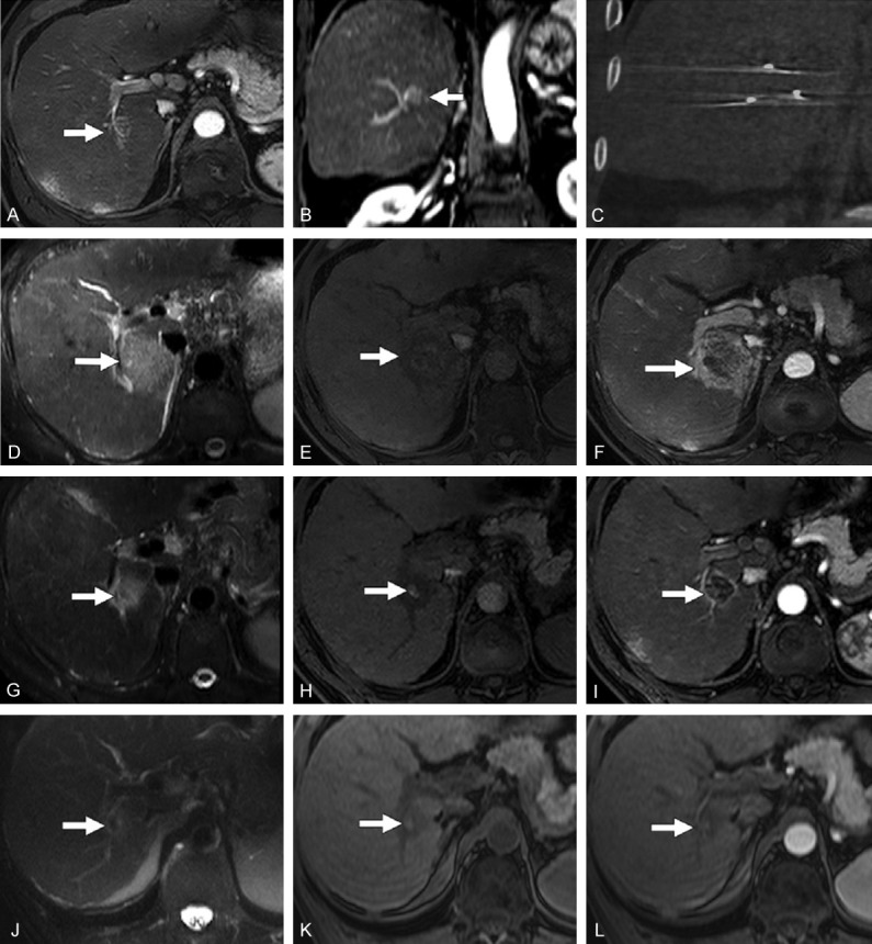
Figure 2.

Typical MRI appearance of HCC after IRE. (A, B) Baseline axial and coronal contrast-enhance T1-weighted MRI acquired in the arterial phase showing an HCC in the right hepatic lobe. (C) Post-IRE CT showing the electrodes. (D-F) Images acquired 24 hours after IRE, showing an ablation area that is hyperintense at T2-weighted MRI (D), slightly hyperintense with an hypointense margin at unenhanced T1-weighted MRI (E) and hypointense with hyperintense margin at contrast-enhanced T1-weighted MRI during the arterial phase (F). (G-I) T2-weighted MRI (G), unenhanced (H) and contrast-enhanced (I) T1-weighted MRI acquired 30 days after IRE, showing similar signals but reduced dimensions with respect to the previous time point. (J-L) T2-weighted MRI (J), unenhanced (K) and contrast-enhanced (L) T1-weighted MRI acquired 120 days after IRE, showing a further reduction of the ablation zone and no contrast enhancement. Reproduced with permission from [19].
