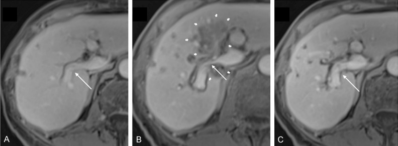
Figure 3.

Portal vein narrowing in a case of colorectal cancer. A: Pre-interventional contrast-enhanced T1-weighted MRI shows a freely perfused right branch of the portal vein (thin arrow). B: 3 days after IRE, MRI shows a caliber reduction of the right portal vein (thin arrow), encased by the ablation zone (thick arrows). C: 6 weeks after treatment, the lumen reduction of the right portal vein (thin arrow) has resolved. Reproduced from [21].
