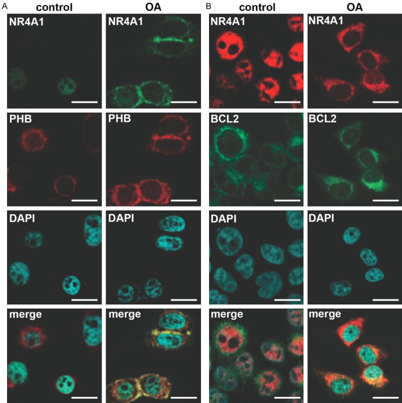
Figure 3.

Localization of NR4A1 in normal and OA patients. A. NR4A1 (green) is translocated to cytoplasm and co-localized with PHB-marked mitochondria (red) in OA patients. B. NR4A1 (red) is translocated to cytoplasm and co-localized with BCL2 (green) in OA patients. Immunohistofluorescence is performed in normal and OA cartilage sections and in each section NR4A1 and PHB or BCL2 are marked with the specific primary antibodies and then the secondary antibodies labelled by Alexa Fluor 647 (red) or 488 (green). DAPI stains nuclei. The experiments are repeated five times. Bar indicates 10 μm. OA, osteoarthritis. NR4A1, nuclear receptor subfamily 4, group A, member 1. PHB, prohibitin. BCL2, B-cell lymphoma 2. merge, pictures showing green, red and blue signals at the same time.
