
Figure 2.
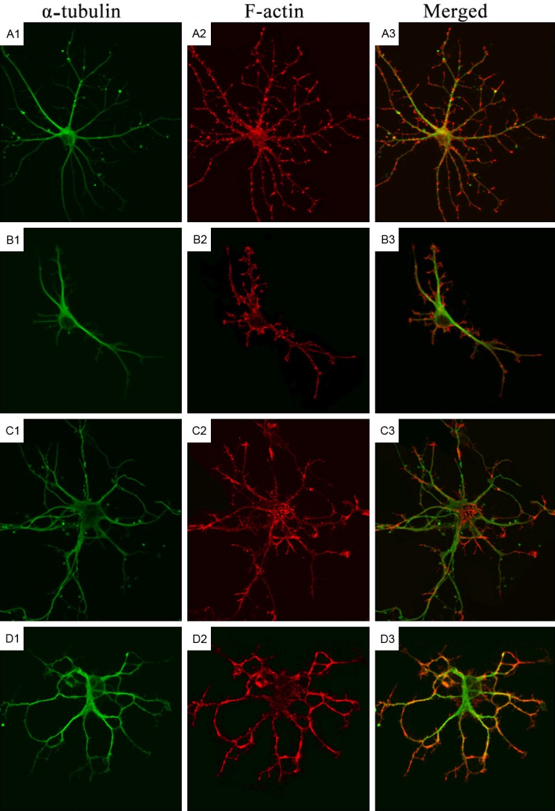
Cytoskeleton of rat hippocampal neurons after treatment with LPA and Y27632 (Confocal Microscopy). A1-A3: Immunofluorescence staining of α-tubulin and F-actin in hippocampal neurons in control group; B1-B3: Immunofluorescence staining of α-tubulin and F-actin in hippocampal neurons in Y-27632 group; C1-C3: Immunofluorescence staining of α-tubulin in hippocampal neurons in LPA group; D1-D3: Immunofluorescence staining of α-tubulin and F-actin in hippocampal neurons in Y-27632+LPA group.