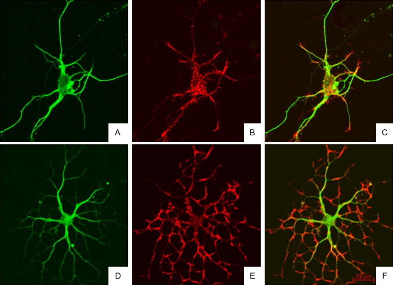
Figure 6.

Cytoskeleton of rat hippocampal neurons after treatment with BAPTA/AM and LPA (Confocal Microscopy). Neurite branches of neurons increased, the microfilaments and microtubules were observed in the neuronal body and protrusions of different levels, and microfilaments were obvious in the protrusions of different levels. A-C: Immunofluorescence staining of α-tubulin and F-actin in hippocampal neurons in BAPTA/AM group; D-F: Immunofluorescence staining of α-tubulin and F-actin in hippocampal neurons in BAPTA/AM+LPA group.
